Knee Osteoarthritis Physiotherapy Protocol — Clinician Version
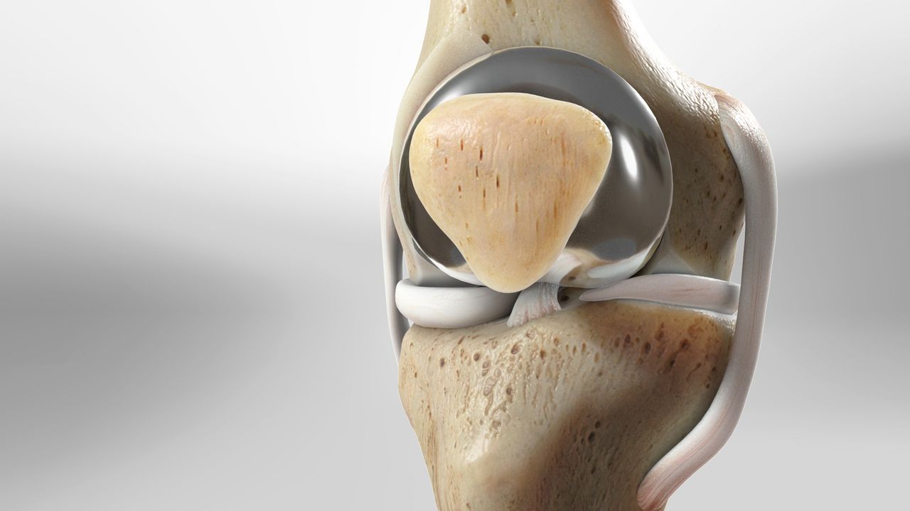
This protocol sets out first-line physiotherapy for symptomatic tibiofemoral and patellofemoral osteoarthritis (OA) in adults.
Purpose and scope
This protocol sets out first-line physiotherapy for symptomatic tibiofemoral and patellofemoral osteoarthritis (OA) in adults. It applies to patients managed non-operatively and to those awaiting arthroplasty or arthroscopic-free pathways. It follows NICE NG226 (2022), OARSI (2019) and the GLA:D and ESCAPE-pain models. It is a framework, not a script. Adjust load, frequency and progression to the patient in front of you. Core principle: exercise is the treatment. Everything else is an adjunct.
- Diagnosis and entry criteria
Clinical diagnosis per NICE. No imaging is required to start treatment. • Age 45 or over • Activity-related knee pain • No morning stiffness, or morning stiffness lasting under 30 minutes Always document the side (right, left or bilateral) and the dominant compartment if known. Record body mass index, comorbidities, and current analgesia. Exclude or escalate before starting • Hot, red, swollen knee with fever or systemic upset — exclude septic arthritis and crystal arthropathy • True locking, giving way with a mechanical block, or acute traumatic effusion • Night pain or rest pain out of proportion, unexplained weight loss, history of malignancy • Inflammatory pattern: polyarticular, prolonged morning stiffness, raised inflammatory markers • Rapidly progressive loss of range or function over weeks
- Baseline assessment
History • Pain: location, NRS at rest and on activity, 24-hour pattern, flare frequency • Function: stairs, sit-to-stand, walking distance, kneeling, work and sport demands • Patient goals — two or three, specific and measurable • Beliefs about the knee: fear of movement, expectation of 'wear and tear', prior advice received • Sleep, mood, weight trajectory, and other joints
Examination
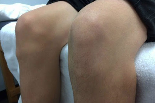
• Gait: antalgic pattern, varus thrust, Trendelenburg • Alignment, effusion (sweep test), warmth, quadriceps bulk • Range of motion with goniometer: flexion, extension, note any fixed flexion deformity • Patellofemoral: crepitus, tracking, apprehension • Strength: quadriceps and hip abductors (MRC grade or hand-held dynamometry if available) • Single-leg balance, single-leg squat quality • Hip and lumbar spine screen to exclude referred pain Outcome measures — record at baseline, 6 weeks, 12 weeks, discharge Domain Measure Minimal clinically important change Patient-reported KOOS (or KOOS-12) — or Oxford Knee Score KOOS subscale 8–10 points; OKS 5 points Pain Numeric Rating Scale, worst pain over 7 days 2 points or 30% Performance 30-second chair stand test 2–3 repetitions Performance 40 m fast-paced walk test 0.2–0.3 m/s Performance Stair climb test (9 or 11 steps, timed) Trend only Performance Timed Up and Go 0.8–1.4 s Global Patient global rating of change (7-point) 'Better' or above
Treatment pillars
4.1 Education and self-management (every patient, every visit)
• OA is a whole-joint condition driven by load, muscle weakness and inflammation. It is not simply 'bone on bone'. Radiographs correlate poorly with symptoms. • Exercise does not damage the joint. Expect some discomfort during and after exercise. Pain up to 5/10 that settles within 24 hours is acceptable. • Set a flare plan on day one (see section 7). • Signpost to Versus Arthritis resources and, where available, a group programme (ESCAPE-pain or GLA:D). • Address unhelpful beliefs directly: 'rest is best', 'avoid stairs', 'wait for the replacement'. 4.2 Exercise therapy (the core intervention) Prescribe three components. Aim for a minimum of 12 supervised sessions over 6–12 weeks, plus home exercise on at least three days per week. Dose matters more than the specific exercise chosen. • Strengthening — quadriceps, gluteus medius and maximus, hamstrings, calf. Start 2–3 sets of 10–15 repetitions at RPE 6–7/10, progressing to 3 sets of 8–12 at RPE 7–8/10. Progress load before volume. • Neuromuscular control — sit-to-stand, step-ups, split squats, single-leg balance, weight-shift and stepping drills. Emphasise knee-over-foot alignment and slow eccentric control. • Aerobic — walking, cycling, aquatic exercise or cross-trainer. Build to 150 minutes of moderate activity per week in bouts of 10 minutes or more. Add range-of-motion work (heel slides, prone hangs, wall slides) where extension or flexion is restricted. A fixed flexion deformity of more than 5° needs daily extension work and a note to the surgeon if it fails to improve. 4.3 Weight management If BMI is 25 or above, discuss weight. A 5–10% loss produces meaningful improvement in pain and function. Refer to Tier 2 or Tier 3 weight services and the GP per local pathway. Do not delay exercise while waiting for weight loss. 4.4 Adjuncts (use with exercise, never instead of it) • Manual therapy: short-term relief only. Use tibiofemoral and patellofemoral mobilisation, soft-tissue work, if it helps the patient engage with exercise. • Heat or cold: patient choice, for symptom relief. • Walking aid: single stick in the contralateral hand if gait is antalgic or the patient has fallen. • Unloader brace or lateral wedged insole for unicompartmental disease with varus or valgus malalignment — trial for 4–6 weeks, keep only if it clearly helps. • Footwear: cushioned, supportive, low heel. Avoid custom orthoses unless there is a clear foot-driven indication. • Topical NSAID: first-line pharmacological adjunct per NICE. Oral NSAIDs with gastroprotection at the lowest dose for the shortest period, via GP. 4.5 Not recommended (NICE NG226) • Acupuncture or dry needling • Electrotherapy: TENS, ultrasound, laser, interferential • Glucosamine, chondroitin, strong opioids • Arthroscopic lavage and debridement for OA alone • Routine intra-articular hyaluronic acid Intra-articular corticosteroid may be considered for a flare that blocks engagement with exercise, discussed with the medical team. Benefit is short-term (2–10 weeks).
- Phased programme
Phases are progression-based. A patient may move through in six weeks or stay in Phase 1 for three months. Progress on criteria, not on the calendar. Phase 1 — Settle and activate (weeks 0–4) Goals: reduce irritability, restore quadriceps activation, establish the exercise habit, agree the flare plan. Component Prescription Quadriceps activation Inner-range quads over a roll, static quads, straight-leg raise — 3 × 10, daily Closed-chain strength Sit-to-stand from raised chair, mini squat to 30–45°, wall sit 20–30 s — 2 × 10, alternate days Hip strength Side-lying abduction, bridge, clam with band — 2 × 12, alternate days Range Heel slides, prone knee hang, seated extension holds — 3 × 30 s, daily Balance Double- to single-leg stance, eyes open, 3 × 30 s, daily Aerobic Walking or stationary cycling, 10–20 minutes, 3–5 days per week, low resistance Education Diagnosis, load tolerance, pain rules, weight, sleep, flare plan Progress when: pain during exercise stays at or below 5/10 and settles within 24 hours; patient completes home programme on 3 or more days per week; sit-to-stand from a standard chair without arms is achievable. Phase 2 — Build strength and control (weeks 4–12) Goals: measurable strength gain, improved chair-stand and walk test scores, return to daily tasks without avoidance. Component Prescription Strength Leg press or goblet squat, step-up (10–20 cm), split squat, Romanian deadlift, calf raise — 3 × 8–12 at RPE 7–8, 2–3 days per week Neuromuscular Step-down with control, lateral step, lunge walk, single-leg squat to box — 3 × 8, quality over depth Hip Banded side-step, single-leg bridge, hip thrust — 3 × 10 Balance / proprioception Single-leg stance on foam, tandem walk, reach tasks — 3 × 45 s Aerobic Build to 30 minutes, 5 days per week, moderate intensity; cycling or aquatic if walking is limiting Range Continue only if a deficit persists Progress when: 30-second chair stand improves by 3 or more repetitions; step-down from 15 cm with good alignment; patient can walk 30 minutes continuously. Phase 3 — Load and function (weeks 12+) Goals: task-specific capacity, return to work, sport or hobby demands, independent long-term programme. Component Prescription Strength Progress load: 3–4 × 6–10 at RPE 8; add unilateral loading. Maintain twice weekly for life Function Task rehearsal: stairs with load, kneeling and floor transfers, carrying, uneven ground, gardening tasks Power (selected patients) Fast sit-to-stand, low box step-up with speed, cycling intervals Aerobic 150 minutes per week moderate, or 75 minutes vigorous, sustained Self-management Written long-term plan, community exercise referral, review of flare plan Discharge when: goals met; KOOS or OKS improved beyond the MCID; patient confident to self-manage flares; independent gym or home programme in place.
- Progression and pain rules
• Traffic light: 0–2/10 green, continue and progress; 3–5/10 amber, continue at the same level; 6/10 or above, or pain not settled by next morning, red — reduce load by a stage. • Increase one variable at a time: load, range, speed, or volume. • Swelling that persists for more than 24 hours after exercise means the dose was too high. Do not stop; regress. • If there is no measurable improvement after 6 weeks of adequate dosing, review adherence, the dose, comorbidities, and consider group programme or medical review.
- Flare management
Give every patient a written flare plan at the first visit.
- Reduce, do not stop. Drop to Phase 1 exercises for 3–5 days.
- Ice or heat for 15 minutes, up to three times daily, whichever the patient prefers.
- Simple analgesia and topical NSAID as agreed with the GP.
- Keep walking short and often rather than one long walk.
- Return to the previous level once pain settles below 5/10 and swelling has resolved.
- Contact the service if the flare lasts more than two weeks, if the knee is hot and red, or if there is a new mechanical symptom.
- Referral triggers for orthopaedic opinion
• Completion of at least 12 weeks of adequately dosed, supervised exercise with no meaningful improvement in pain or function • Night pain that disturbs sleep despite optimised non-operative care • Progressive deformity, fixed flexion deformity over 10°, or instability • Severe limitation of walking distance (under 500 m) or stairs despite a full programme • Patient has considered and wishes to discuss surgical options Include in the referral: side, duration, outcome scores at baseline and latest, exercise programme delivered, weight trajectory, walking distance, and patient expectations. Patients on the surgical pathway should continue Phase 2 strengthening pre-operatively; quadriceps strength predicts early post-operative recovery.
- Documentation standard
• Side and compartment stated in every entry • Outcome measures at the intervals in section 3 • Exercise prescription with sets, repetitions, load and RPE • Adherence recorded as days per week completed • Flare plan given and understood — documented once • Weight and BMI at baseline and discharge
- Key references
- NICE. Osteoarthritis in over 16s: diagnosis and management. NG226. 2022.
- Bannuru RR et al. OARSI guidelines for the non-surgical management of knee, hip, and polyarticular osteoarthritis. Osteoarthritis Cartilage 2019;27:1578–89.
- Fransen M et al. Exercise for osteoarthritis of the knee. Cochrane Database Syst Rev 2015;1:CD004376.
- Skou ST, Roos EM. Good Life with osteoArthritis in Denmark (GLA:D). BMC Musculoskelet Disord 2017;18:72.
- Hurley MV et al. Long-term outcomes and costs of an integrated rehabilitation program for chronic knee pain (ESCAPE-pain). Arthritis Rheum 2007;57:1220–9.
- Messier SP et al. Effects of intensive diet and exercise on knee joint loads, inflammation, and clinical outcomes (IDEA). JAMA 2013;310:1263–73.
- Dobson F et al. OARSI recommended performance-based tests to assess physical function in people diagnosed with hip or knee osteoarthritis. Osteoarthritis Cartilage 2013;21:1042–52.